Home
/ Back Of Knee Anatomy - Anterior And Posterior Aspects Of The Knee Netter Shoulder Anatomy Knee Joint Anatomy Neck And Shoulder Muscles : It may be caused by another problem, like arthritis or a tear in your.
Back Of Knee Anatomy - Anterior And Posterior Aspects Of The Knee Netter Shoulder Anatomy Knee Joint Anatomy Neck And Shoulder Muscles : It may be caused by another problem, like arthritis or a tear in your.
Back Of Knee Anatomy - Anterior And Posterior Aspects Of The Knee Netter Shoulder Anatomy Knee Joint Anatomy Neck And Shoulder Muscles : It may be caused by another problem, like arthritis or a tear in your.. By stretching and strengthening the knee muscles, you can reduce the forces going through the knee joint, reducing pain and swelling, and improving function. Do you mean the back view if yes then it is called the posterior view of the knee. Articular surface of the femur. The hamstrings start at the pelvis, cross the back of the knee, and attach to your tibia or shin bone. These four ligaments act collectively to support and stabilize the knee throughout its range of motion.
Ebraheim's educational animated video describes the anatomy of the knee joint.•femur •tibia•fibula•patella•joint capsule: A popliteal aneurysm is where there is an abnormal bulge in the popliteal artery, the main artery at the back of the knee. The distinguishing feature of a popliteal aneurysm is a palpable pulsating mass behind the knee. The knee is one of the biggest and most complex joints in the human body. The knee consists of three bones:
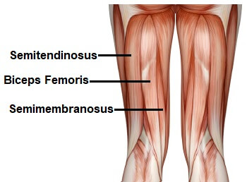
Clinical Anatomy Of The Knee Reumatologia Clinica from multimedia.elsevier.es It's often linked to other conditions affecting the knee, including osteoarthritis, rheumatoid arthritis, cartilage injuries and inflammation of the knee joint. Anatomy of the back of the knee in order to better understand what you're stretching when performing a hamstring stretch let's talk about what's back there. A hamstring tendon strain is a tear of one of the hamstring tendons. In addition to reading this article, be sure to watch our knee anatomy animated tutorial video. The distinguishing feature of a popliteal aneurysm is a palpable pulsating mass behind the knee. The back of the knee is a complicated area. The term anterior refers to the front of the knee, while the term posterior refers to the back of the knee. The side that is away from midline if called lateral side of the knee and the closer.
A cyst is a collection of fluid inside a thin layer.
The distinguishing feature of a popliteal aneurysm is a palpable pulsating mass behind the knee. So the anterior cruciate ligament is in front of the posterior cruciate ligament. The knee joint is part of the lower extremity. The knee consists of three bones: These four ligaments act collectively to support and stabilize the knee throughout its range of motion. The term anterior refers to the front of the knee, while the term posterior refers to the back of the knee. If you think about it, your knee supports almost all of your body weight, which is a big job in itself. It may be caused by another problem, like arthritis or a tear in your. From a functional perspective, we have many muscles, such as the hamstring and calf muscles. The hamstrings start at the pelvis, cross the back of the knee, and attach to your tibia or shin bone. It is more common in sprinters or sports involving kicking. A cyst is a collection of fluid inside a thin layer. By stretching and strengthening the knee muscles, you can reduce the forces going through the knee joint, reducing pain and swelling, and improving function.
It is the junction of the thigh and the leg and is a hinge joint. In this video, i explain important anatomy of knee joint. Back of the knee anatomy. The distinguishing feature of a popliteal aneurysm is a palpable pulsating mass behind the knee. There are several critical structures back there.
The Knee Anatomy Injuries Treatment And Rehabilitation from i0.wp.com Normal anatomy and biomechanics of the knee fred flandry, md, facs*w and gabriel hommel, md* abstract: The hamstring muscles are three muscles at the back of the thigh that affect hip and knee movement. The ligament that gives stability to the inner knee. Ebraheim's educational animated video describes the anatomy of the knee joint.•femur •tibia•fibula•patella•joint capsule: A popliteal cyst is a cyst in the shallow depression at the back of the knee. If you think about it, your knee supports almost all of your body weight, which is a big job in itself. A special characteristic of the knee that differentiates it from other hinge joints is that it allows a small degree of medial and lateral rotation when it is moderately flexed. The knee is the joint where the bones of the lower and upper legs meet.
The knee connects the thigh bone (femur located in the upper leg) to the shinebone (tibia located in the lower leg.the calf bone (fibula located in the lower leg) connects to the joint, but is not directly affected by the hinge joint action.
Our knee anatomy is comprised of bones (the tibia also known as the shin bone, the fibula which is the smaller bone that runs alongside the tibia, the thigh bone also known as the femur, and the patella which is also known as the kneecap), tendons, muscles, cartilage and ligaments (the acl, pcl, lcl and mcl). Functionally, the knee comprises 2 articulations—the patellofemoral and tibiofemoral. The knee joint is part of the lower extremity. The side that is away from midline if called lateral side of the knee and the closer. It may be caused by another problem, like arthritis or a tear in your. In addition, pathological swelling of this portion of the knee, which usually occurs due to a leak in the posteromedial joint capsule, is called a baker's cyst. It is more common in sprinters or sports involving kicking. To put it plainly, sometimes hip pain comes from the hip, but a lot of times hip pain comes from the back. The knee joins the thigh bone (femur) to the shin bone (tibia). Things being what they are, your knee underpins the majority of your body weight, which is a difficult task in itself. Learn more about causes, how to treat it, and outlook here. This guide will help you understand Anatomy of the back of the knee in order to better understand what you're stretching when performing a hamstring stretch let's talk about what's back there.
The knee connects the thigh bone (femur located in the upper leg) to the shinebone (tibia located in the lower leg.the calf bone (fibula located in the lower leg) connects to the joint, but is not directly affected by the hinge joint action. The distinguishing feature of a popliteal aneurysm is a palpable pulsating mass behind the knee. The hamstring muscles are three muscles at the back of the thigh that affect hip and knee movement. A popliteal cyst is a cyst in the shallow depression at the back of the knee. Many types of injury can cause a collection of fluid.
Knee Muscles Anatomy Function Injuries Knee Pain Explained from www.knee-pain-explained.com The knee joint is part of the lower extremity. Pain in the back of the knee has many potential causes, including baker's cysts and muscle injuries. The knee is a complex joint that has many functions. Anatomy muscles of the knee, muscle anatomy back of knee, muscle anatomy behind the knee, muscle anatomy of back of knee, human muscles, anatomy muscles of the knee. In addition to reading this article, be sure to watch our knee anatomy animated tutorial video. It's often linked to other conditions affecting the knee, including osteoarthritis, rheumatoid arthritis, cartilage injuries and inflammation of the knee joint. They begin under the gluteus maximus behind the hip bone and attach to the tibia at the knee. Many types of injury can cause a collection of fluid.
The knee joint is a synovial joint which connects the femur (thigh bone), the longest bone in the body, to the tibia (shin bone).
Normal anatomy and biomechanics of the knee fred flandry, md, facs*w and gabriel hommel, md* abstract: A popliteal cyst is a cyst in the shallow depression at the back of the knee. If you think about it, your knee supports almost all of your body weight, which is a big job in itself. The side that is away from midline if called lateral side of the knee and the closer. The knee is the joint where the bones of the lower and upper legs meet. They begin under the gluteus maximus behind the hip bone and attach to the tibia at the knee. 1) the tibiofemoral joint where the tibia meet the femur 2) the patellofemoral joint. The ligament, located in the back of the knee, that controls backward movement of the tibia (shin bone). The term anterior refers to the front of the knee, while the term posterior refers to the back of the knee. Popliteal aneurysms tend to be asymptomatic and are the most common cause of swelling behind the knee with no pain. Back of knee anatomy the knee is one of the largest and most complex joints in the human body. Anatomy muscles of the knee, muscle anatomy back of knee, muscle anatomy behind the knee, muscle anatomy of back of knee, human muscles, anatomy muscles of the knee. The largest joint in the body, the knee moves like a hinge, allowing you to sit, squat, walk or jump.